Hidden decay treated with the dental microscope
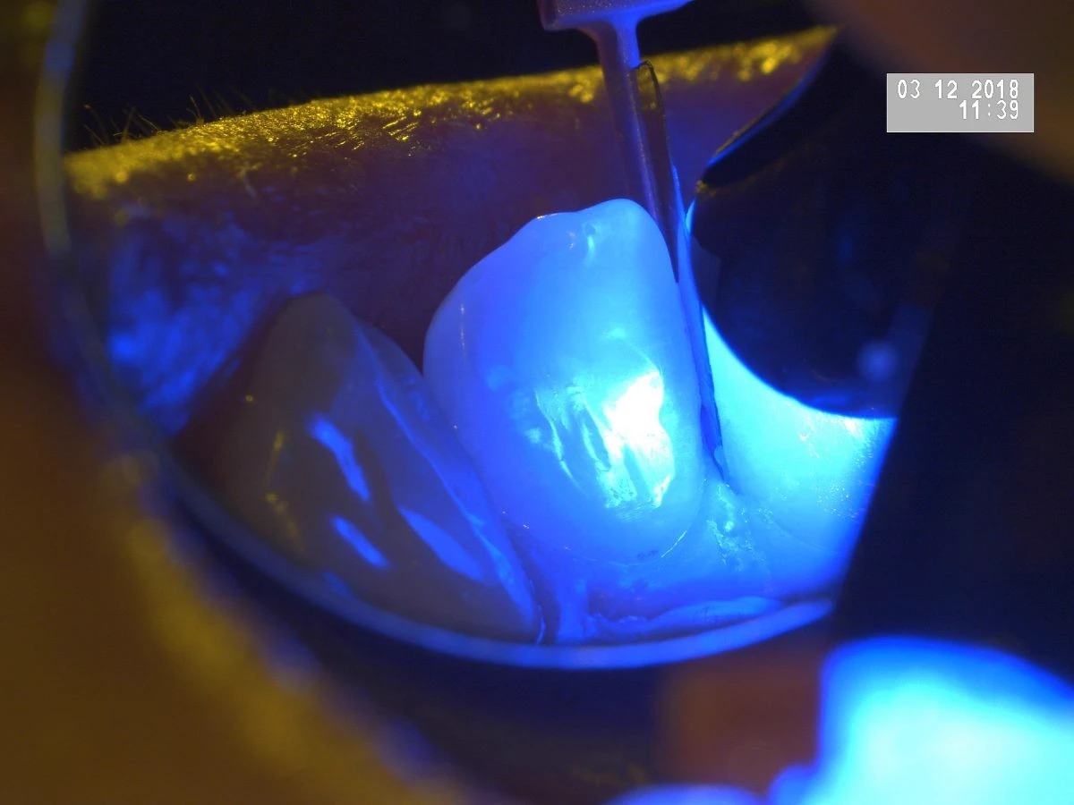
Tooth decay often develops without clinical signs (it doesn’t hurt). That is why a regular dental check-up every 6 months is essential for detecting it early. Our high-performance Leica dental microscope gives us precise magnification for the most accurate diagnosis possible.
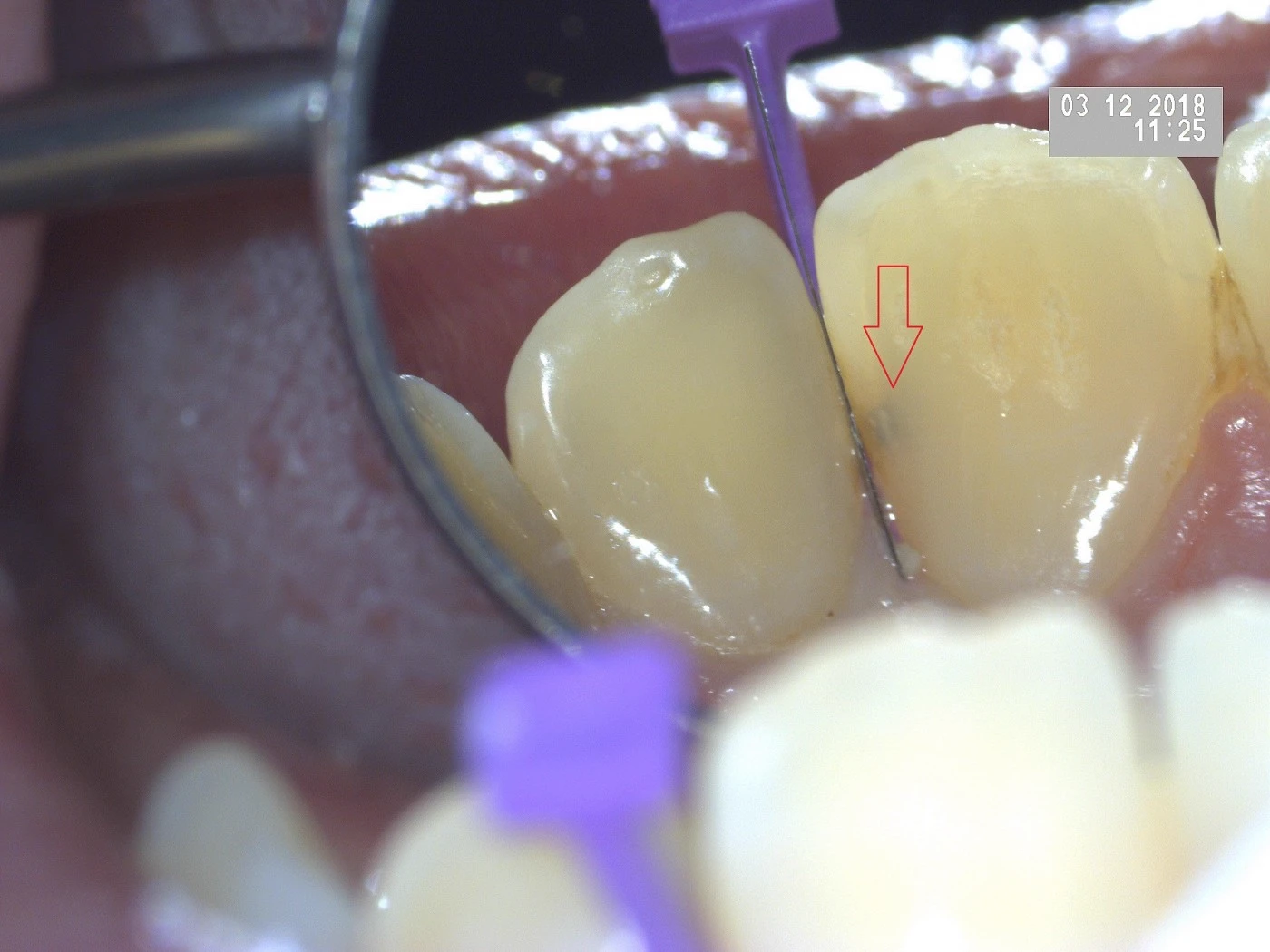
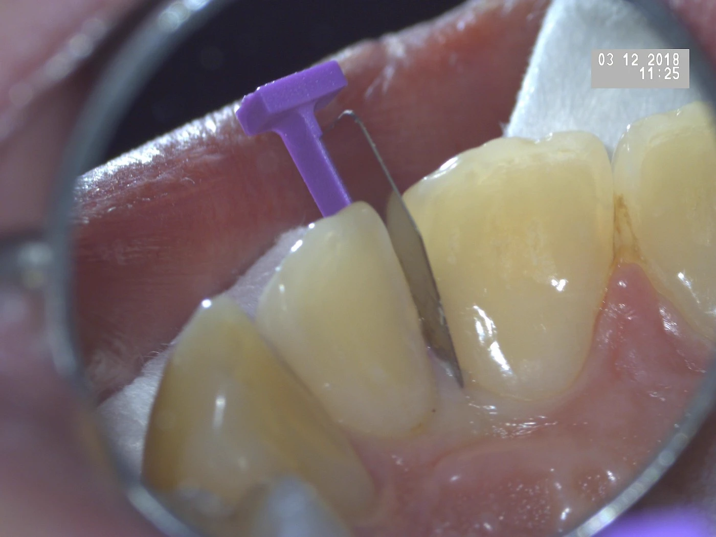
To protect the neighbouring tooth, we use special protective devices such as this metal separator.
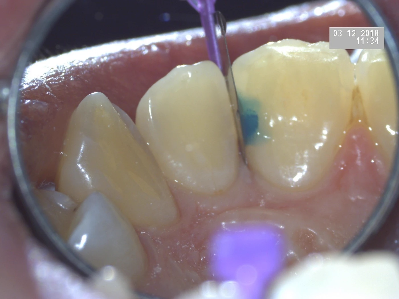
Once access has been created, you can see how large the decay actually is.
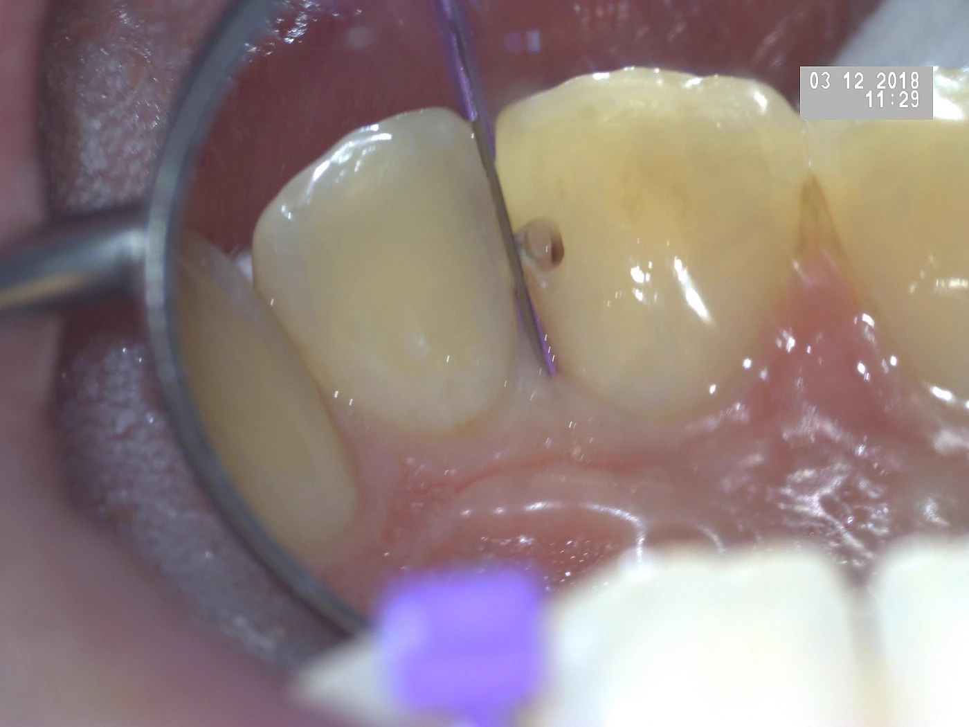
Because the decay was detected in time, we were able to work without anaesthetic. The decay was removed completely using dental burs.
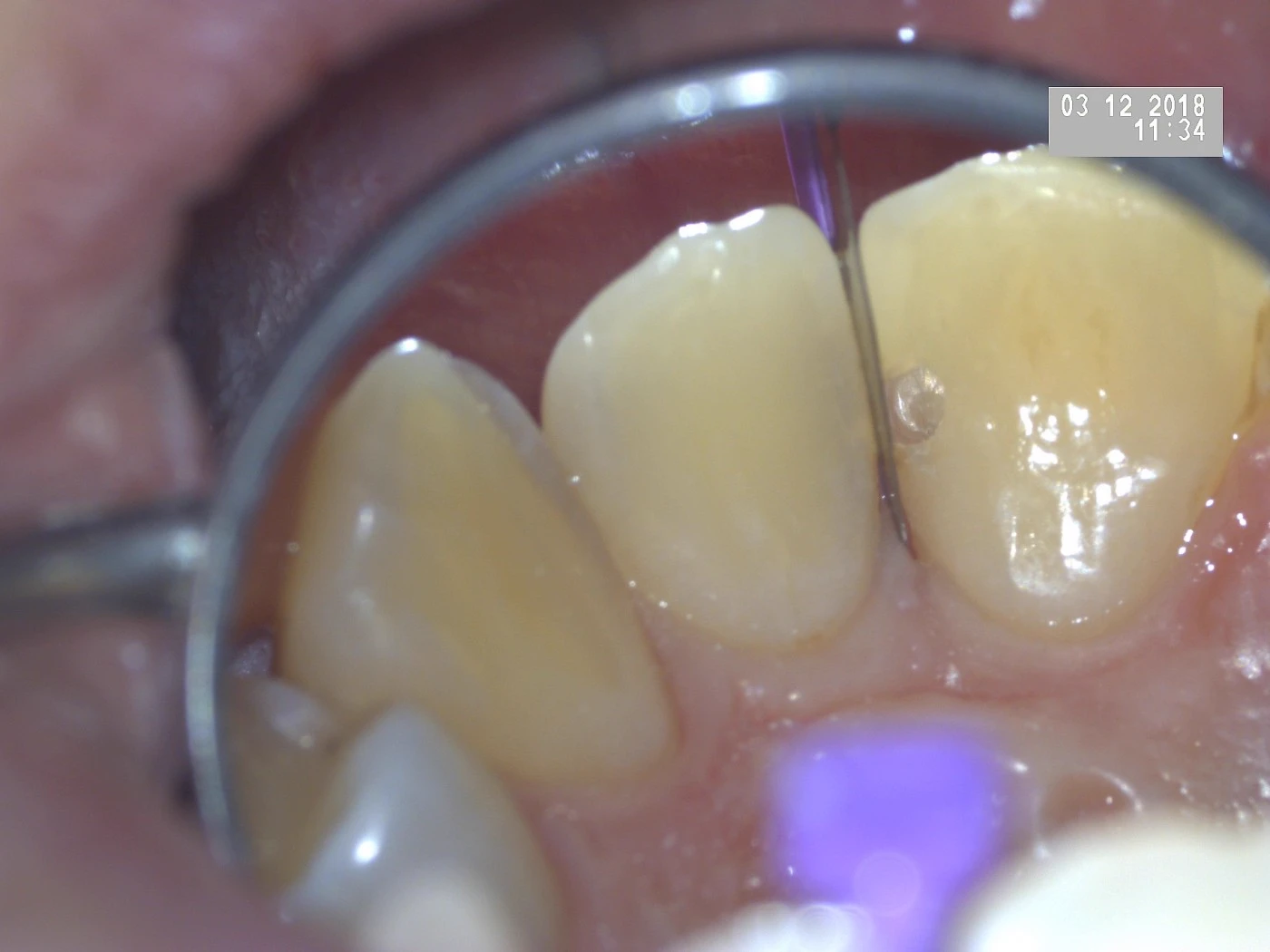
The surface of the tooth is conditioned so that an aesthetic ‘filling’ can be applied.
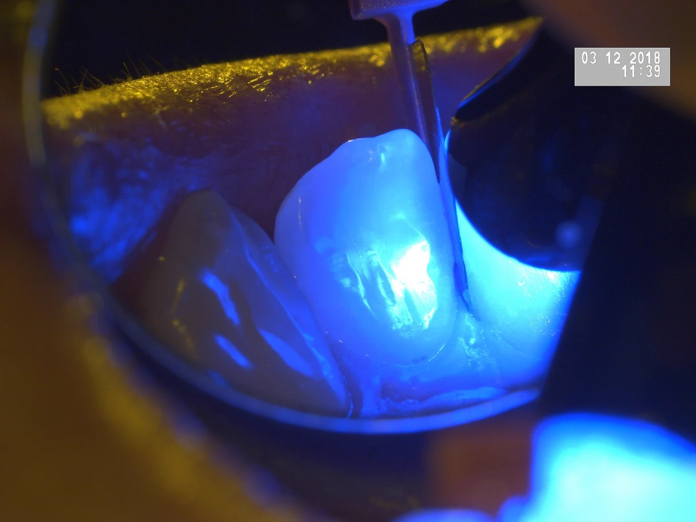
The aesthetic filling material is applied and cured with the LASER lamp.
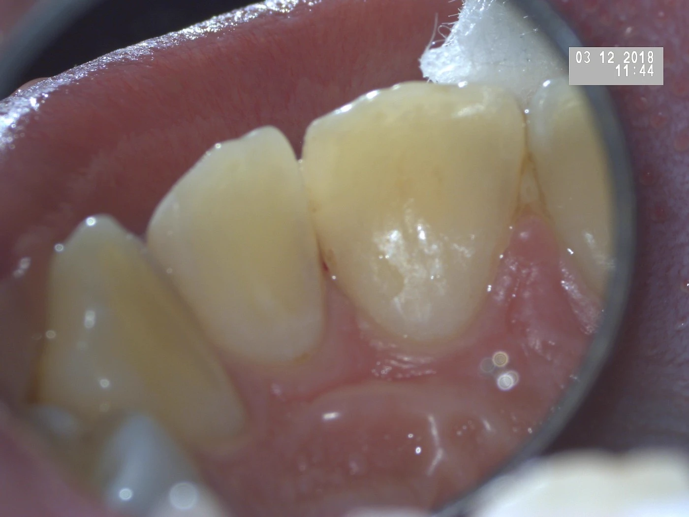
The final result, achieved in less than 20 minutes.
Lasă-ne un comentariu